When it comes to comprehensive dental and orthodontic treatment planning, few diagnostic tools are as valuable as the cephalometric X-ray. This sophisticated imaging technique provides dentists and orthodontists with crucial information about your facial structure, helping them develop the most effective treatment plans for various dental and orthodontic concerns.
What is a Cephalometric X-ray?
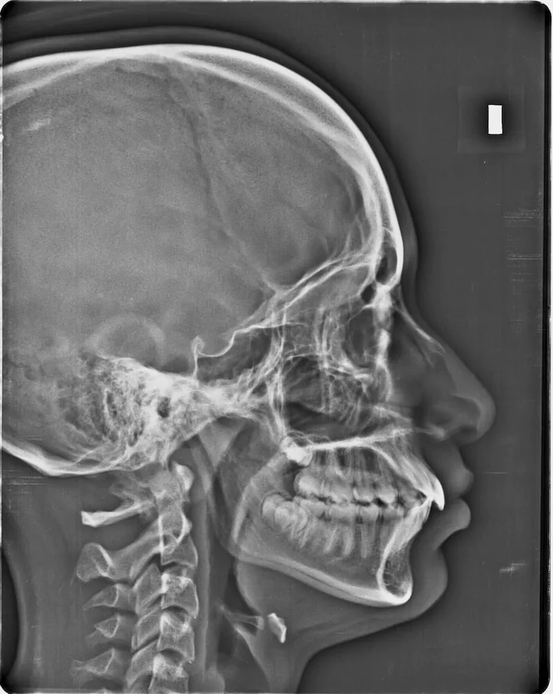
A cephalometric X-ray, often called a "ceph" in dental circles, is a specialized lateral (side-view) X-ray of the skull and facial bones. Unlike traditional dental X-rays that focus solely on the teeth, cephalometric X-rays capture the entire side profile of your head, including the skull, facial bones, soft tissue profile, and airway spaces.
How Does it Work?
The process involves positioning the patient's head in a specialized cephalometric machine, which maintains a standardized distance between the X-ray source and the patient's head. This consistency is crucial for accurate measurements and comparisons over time. The X-ray beam passes through one side of the head and is captured on the opposite side, creating a detailed two-dimensional image.
Key Information Revealed by Cephalometric X-rays:
- Skeletal Relationships
- Position and size of the upper and lower jaws
- Growth patterns of facial bones
- Relationship between the skull base and jaw structures
- Vertical facial proportions
- Dental Characteristics
- Position and inclination of front teeth
- Relationship between teeth and supporting bone
- Dental crowding or spacing issues
- Soft Tissue Profile
- Lip position and thickness
- Nose projection
- Chin prominence
- Overall facial balance
- Airway Analysis
- Size and shape of the airway passage
- Potential breathing obstruction issues
- Adenoid tissue evaluation
Clinical Applications
Orthodontic Planning: Cephalometric X-rays are invaluable for orthodontic treatment planning. They help orthodontists:
- Predict growth patterns in younger patients
- Determine the best approach for bite correction
- Plan tooth movement strategies
- Evaluate the need for surgical intervention
- Monitor treatment progress
Surgical Planning: For patients requiring orthognathic surgery (jaw surgery), cephalometric X-rays provide essential information for:
- Surgical approach determination
- Precise measurement of necessary corrections
- Post-surgical outcome prediction
- Treatment simulation
Sleep Apnea Assessment: The airway analysis capabilities make cephalometric X-rays useful in:
- Evaluating airway obstruction
- Planning sleep apnea treatments
- Monitoring changes in airway dimensions
Safety Considerations
While cephalometric X-rays do involve radiation exposure, the dose is minimal and carefully controlled. Modern digital X-ray equipment uses significantly less radiation than traditional film X-rays. The benefits of the diagnostic information obtained generally outweigh the minimal risks associated with radiation exposure.
The Technology Behind the Images
Modern cephalometric analysis has gone digital, offering several advantages:
- Instant image availability
- Enhanced image quality
- Digital storage and easy sharing
- Reduced radiation exposure
- Advanced analysis software capabilities
Treatment Planning and Patient Communication
One of the most valuable aspects of cephalometric X-rays is their role in patient education and treatment planning:
- Visual demonstration of existing conditions
- Clear communication of treatment goals
- Progress monitoring and documentation
- Treatment outcome prediction
- Before-and-after comparisons
When Are Cephalometric X-rays Needed?
These X-rays are typically recommended:
- Before beginning orthodontic treatment
- When evaluating growth and development
- For surgical treatment planning
- When assessing airway problems
- For monitoring treatment progress
The Future of Cephalometric Analysis
The field continues to evolve with:
- 3D cephalometric analysis capabilities
- Artificial intelligence-assisted analysis
- Enhanced diagnostic accuracy
- Improved treatment prediction models
- Integration with other imaging modalities
Conclusion
Cephalometric X-rays represent a cornerstone of modern dental and orthodontic diagnosis and treatment planning. Their ability to provide detailed information about skeletal, dental, and soft tissue relationships makes them an invaluable tool in achieving optimal treatment outcomes. As technology continues to advance, the applications and capabilities of cephalometric analysis will only continue to expand, further improving patient care and treatment precision.
Remember, while this information provides a comprehensive overview of cephalometric X-rays, each patient's needs are unique. Consult with your dental professional to understand how this diagnostic tool might benefit your specific treatment plan.